

Use this ACLS exam ECG rhythm identification guide to build a reading routine you can explain under pressure. Start with the patient description, describe the tracing, and connect each answer to a visible finding. Keep your current ACLS course materials beside you to verify diagnostic criteria, reference rhythms, and algorithm choices.
The study goal is specific: give a rhythm interpretation, identify the evidence behind it, and recognize which information you still need. Practice that reasoning before adding a timer. For clinical assessment and treatment, follow your current training and local protocols.
Keep the patient description beside the tracing
For every practice question, separate what you can observe on the ECG from what the scenario tells you about the patient. Write those findings on different lines before choosing an answer.
On the tracing line, record rate, regularity, identifiable complexes, atrial activity, and the relationship between atrial and ventricular activity. On the patient line, record the stated pulse assessment, symptoms, responsiveness, and other supplied findings.
Avoid filling gaps with assumptions. If the question supplies pulse status, use it explicitly. If it leaves that information unstated, identify the uncertainty before assigning a label that depends on it.
Match your answer to the question
Underline the task before studying the answer choices. Is the question asking for a rhythm name, an assessment, or an action?
For a rhythm question, explain the electrical pattern. For a management question, consult the appropriate course algorithm and include the patient information that determines the pathway.
Use this sentence during review:
“My interpretation is ___ because the tracing shows ___, and the scenario states ___.”
Leave an element blank when the evidence is missing. Then ask whether the question actually requires that element.
Rehearse the handoff separately
After interpreting a strip, practice a brief verbal handoff: your observations, your interpretation, and the relevant patient findings. Keep uncertainty explicit.
Use the ACLS megacode testing checklist as a separate study prompt for rehearsing how you communicate those findings during a scenario. Check clinical decisions against your course materials rather than treating a study checklist as the clinical authority.
Read each practice strip in a consistent order
Choose an observation sequence and use it throughout a study session. Adjust the sequence during review if you discover a recurring omission; avoid changing it halfway through every difficult question.
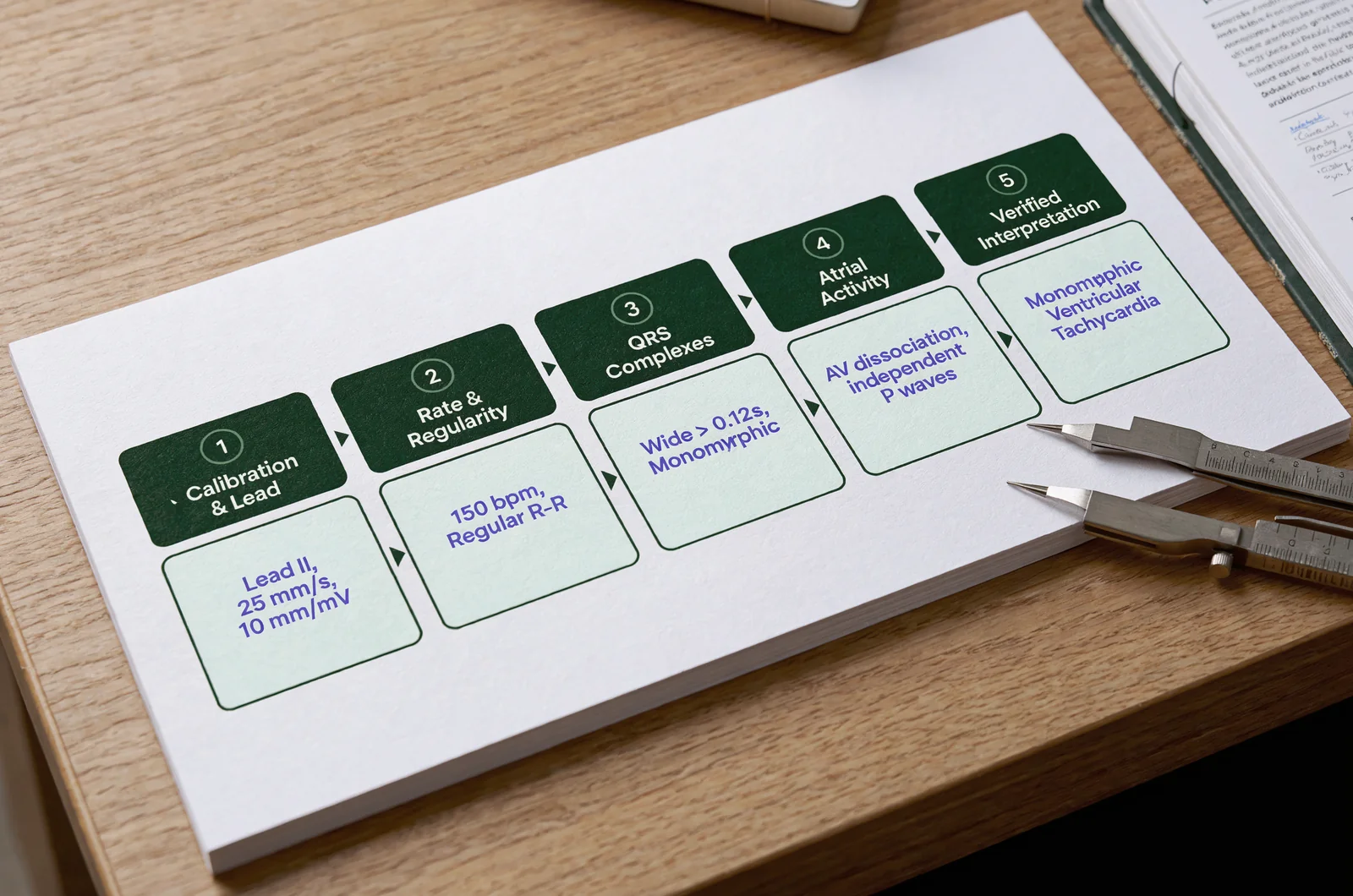
I recommend the following sequence for deliberate practice. Treat it as a study routine, rather than a reason to delay assessment or resuscitation.
Check the tracing before measuring it
Begin with the information attached to the strip. Look for the displayed lead, recording speed, calibration, and available duration.
Confirm which measurements the question permits. Avoid assigning precise intervals to an image whose scale you cannot establish.
Inspect the baseline and the full recording. If part of the tracing is obscured, describe that limitation. Where the exercise supplies another lead or recording, compare it before treating an ambiguous appearance as settled.
For your notes, use concrete language: “The baseline is difficult to follow in the middle of the strip.” Avoid replacing an observation with an unsupported explanation.
Describe rate and regularity
Estimate the rate with a method appropriate to the displayed recording. Verify the calculation method and reference ranges in your course materials.
Then examine the spacing between successive ventricular complexes across the entire strip. Describe whether the spacing appears consistent, follows a repeating variation, or varies without an obvious repeating pattern.
Keep the description separate from the rhythm name. Write what you measured before deciding what it means.
If you change your interpretation after reviewing the answer, retain the original observation. Correct the observation only when you can identify a measurement or reading error.
Examine ventricular complexes
Identify the beginning and end of representative QRS complexes. Compare their duration with the threshold specified in your course materials.
Inspect several complexes rather than relying on the clearest beat alone. Note whether their shapes remain similar or change across the recording.
Before assigning a rhythm from a familiar silhouette, explain which observations support your choice. Require more evidence than speed or appearance alone when distinguishing between plausible alternatives.
Inspect atrial activity and its relationship to the ventricles
Look deliberately for atrial activity. Where you can identify it, examine its timing, appearance, and relationship to ventricular complexes.
Ask whether you can follow the atrial pattern through the full strip. Check the relevant intervals and any apparent interruptions in conduction against your reference examples.
When atrial activity is unclear, write “not clearly identified.” Keep that uncertainty distinct from a confident statement that it is absent.
Finish with a plain-language description before naming the rhythm: rate estimate, regularity, ventricular appearance, atrial findings, and their relationship.
Compare VF with ventricular tachycardia
Study ventricular fibrillation and ventricular tachycardia with verified course examples beside you. Build your comparison around observable electrical features, then add the patient information needed for the scenario.
For each example, explain its position in your current cardiac arrest or tachycardia algorithm. Verify that classification directly rather than relying on a remembered list.
Examine the organization of the tracing
For a course example labeled ventricular fibrillation, ask whether you can consistently identify individual ventricular complexes. Examine the activity throughout the recording, including sections where its amplitude changes.
Write down the visual evidence used in the reference explanation. Compare that evidence with a verified ventricular tachycardia example.
For the VT example, examine the repeated complexes, their spacing, and their shape. Describe how confidently you can follow successive ventricular events.
Avoid using “looks chaotic” or “looks fast” as your complete explanation. Replace each phrase with the particular observation that supports it.
Keep the pulse qualifier explicit
When a question asks about pulseless ventricular tachycardia, identify where the scenario establishes pulse status. Require that information before adding the qualifier to your answer.
For a VT example with a stated pulse, reread the question before deciding which algorithm applies. Include the supplied symptoms and assessment findings in that review.
Practice explaining which part of your conclusion comes from the tracing and which part comes from the patient description. Keep those explanations separate in your answer notes.
Add the mechanism after the visual description
Use your course reference to explain the electrical mechanism behind each verified example. Ask how that explanation accounts for the appearance you described.
Write the mechanism in your own words, then compare it with the reference. Correct terminology that changes the meaning.
End each review with two statements: the observation that supports your answer and the observation that would make you reconsider it.
Verify the evidence behind PEA and asystole answers
For pulseless electrical activity and asystole exercises, make the electrical findings and the patient assessment explicit. Verify the definitions and algorithm placement in your current course materials before building timed drills.
Use a separate review page for each label. Record the reference definition, the evidence required by that definition, and an example of an answer that lacks enough supporting information.
Require the full scenario for a PEA interpretation
When reviewing a course example labeled PEA, identify the electrical description and the stated pulse assessment. Explain how the reference combines those findings.
Avoid memorizing the example’s shape as your entire recognition rule. Instead, compare several verified examples and identify the patient information used in each explanation.
During ExamJungle study sessions, write down the scenario detail that supports your interpretation before reviewing the answer. Afterward, check the clinical reasoning against your current course reference.
If your answer depends on information the question never supplied, mark that assumption in your notes.
Scrutinize a nearly flat tracing
Before selecting asystole in a practice question, inspect the full recording and any information about leads, connections, or recording quality.
Compare the example with the confirmation steps taught in your course. Identify which checks the scenario has already completed and which findings remain uncertain.
Where your materials compare asystole with fine ventricular fibrillation or a recording problem, study those examples together. Describe the distinguishing evidence in each explanation.
Avoid treating a nearly flat image as sufficient justification on its own. State what the exercise establishes about the recording and the patient.
Connect the label to the correct reference pathway
After identifying either rhythm in a verified exercise, locate it in the applicable course algorithm. Explain why that pathway fits the complete scenario.
Keep algorithm review separate from waveform recognition in your notes. Require a supported answer for both before marking the exercise complete.
Give atrial rhythms and AV blocks their own evidence
Build separate comparison sets for atrial rhythms, sinus rhythms, and atrioventricular conduction patterns. Use the same observation sequence, but choose comparison questions that suit each group.
Avoid collecting isolated screenshots without explanations. Beside every verified example, record the diagnostic criteria from your reference and identify where the tracing demonstrates them.
Compare atrial fibrillation and atrial flutter deliberately
Place labeled course examples beside each other. Examine ventricular spacing, atrial activity, and the relationship between the two.
For each example, answer three questions:
- What atrial activity can I identify?
- How would I describe ventricular regularity?
- Which reference criterion supports this interpretation?
Include examples with different appearances when your materials provide them. Recheck your reasoning whenever your answer depends on one familiar-looking segment.
Before moving on, explain why the closest alternative fits less well.
Make sinus-rhythm explanations specific
For a proposed sinus-rhythm interpretation, identify the atrial findings and their relationship to the ventricular complexes. Measure the relevant intervals where the image permits.
Verify the rate category against your course reference. Keep that rate category separate from the evidence supporting the rhythm’s origin.
In a scenario that asks about management, return to the supplied patient assessment after naming the rhythm. Use the applicable algorithm to check what additional information matters.
Trace atrial and ventricular events through AV-block examples
For each verified atrioventricular block example, mark the atrial events and ventricular complexes separately. Follow their timing across the strip.
Inspect the relevant intervals, any repeating sequences, and any apparent interruptions. For advanced-block examples, examine the relationship between atrial and ventricular activity across the full recording.
Compare your marks with the reference explanation before memorizing the name. When your interpretation differs, identify the specific event or interval that accounts for the disagreement.
Repeat the exercise on a different verified example before considering that distinction settled.
Rehearse the details that would change your answer
Use hypothetical variations to test your reasoning after studying verified examples. Label these variations as practice exercises and check every resulting interpretation against your course materials.
Change one detail at a time. Keep the remaining observations fixed so you can explain exactly why you reconsidered the answer.
Change the patient information
Imagine a practice vignette with a rapid, broad-complex tracing. In one version, the scenario states that a pulse is present. In another, it explicitly states that no pulse is detected.
Describe the electrical observations in each version. Then identify which interpretation or algorithm decision requires the changed patient information.
Avoid inventing additional symptoms to make an answer fit. Work only with the stated findings, and record any further assessment the question requires.
Extend the exercise by changing the task: first ask for the rhythm description, then ask which course algorithm should guide the response.
Change the confidence in the recording
Imagine a separate practice exercise with a difficult-to-interpret tracing. Compare a version that supplies another clear lead with one that provides no additional recording.
Write what you can conclude in each version. Identify which uncertainty the additional information resolves.
Check your proposed confirmation steps against your course materials. Avoid repeatedly inspecting the same unclear segment without stating what evidence you need.
Defend the closest alternative
For every difficult question, select the most plausible competing answer. Explain what finding would support it and what finding argues against it in the supplied exercise.
If you cannot distinguish the alternatives, return to paired reference examples. Write a comparison based on observations before attempting another timed question.
Build a practice routine around explanations
Organize practice around a defined task: accurate observations, rhythm interpretation, scenario integration, or algorithm review. Choose the task before opening the question set.
Begin with untimed exercises and require a written explanation. Add timing after you can complete the observation sequence without omitting its essential parts.
Record the reason behind each correction
For an incorrect or uncertain answer, write down your original choice, the finding you used, and the finding you overlooked or misread.
Classify the correction precisely. Did you measure an interval incorrectly? Miss a stated pulse assessment? Choose a label before inspecting atrial activity?
Use the correction to choose the next exercise. For a measurement problem, return to labeled measurement examples. For a scenario-reading problem, underline the supplied patient findings before examining the next tracing.
Keep correctly answered questions in review when your reasoning remains uncertain.
Compare explanations with a clinical reference
If you include ExamJungle in your practice routine, keep a separate clinical verification step. Compare diagnostic explanations and algorithm choices with your current ACLS materials.
When explanations disagree, identify the exact disputed claim. Bring that question to your instructor or another appropriate clinical educator rather than resolving it by counting repeated answers.
Record the reference used to settle the question. Update the affected study note before repeating the exercise.
Use an interactive worksheet to expose missing observations
A useful study tool would present a verified tracing and require separate entries for regularity, ventricular appearance, atrial findings, and supplied patient information before accepting a rhythm choice.
Its feedback should connect each correction to an approved clinical reference. Include a field for uncertainty so learners can distinguish an observation they missed from information the exercise never supplied.
For your final rehearsal, choose previously reviewed examples in a new order. Explain each answer before checking the reference.
Resolve uncertainty before the next timed set
Use the questions below to decide what to review when a rhythm exercise leaves you uncertain. Keep the response tied to the specific evidence you need.
Should I memorize numerical thresholds first?
Learn the thresholds specified in your current course, then practice measuring them on verified examples. Pair each number with the feature being measured and the interpretation it supports.
Avoid adding remembered values to your notes without checking their definitions and context.
What should I do when two answers still seem plausible?
Write the single finding that would distinguish them. Search the supplied tracing and vignette for that finding, then review the reference explanation.
If the evidence remains insufficient, record the ambiguity and ask your instructor to clarify the exercise. Keep a forced guess separate from a supported interpretation.
How should I review an answer I got right by guessing?
Rework it without the choices. Describe the tracing, identify the relevant patient findings, and justify the interpretation.
Then compare it with the closest alternative. Retain the question for review until you can explain both the correct choice and the rejected option.
How should I judge my readiness?
Use a review standard that requires a defensible explanation for each answer. Include unfamiliar-looking verified examples, scenario changes, and questions that require you to identify missing information.
Before ending the session, record the exact observation or reference you need to review next.
Continue with scenario and emergency exam practice